Computed tomography

Location: Braunschweig (headquarters)

© Fraunhofer WKI
Computer tomograph

Why has a component failed? Is the cause of the problem in the material or in the manufacturing process? How can component properties such as dimensional accuracy or strength be optimized? We find out for you. Using computed tomography, we examine materials and components non-destructively regarding quality characteristics and damage. In this way, we support you very efficiently in the new and further development of materials, components and production processes as well as in quality assurance.

Computed tomography (CT) enables a three-dimensional imaging of the internal and external structure of objects with a detail detectability down into the micrometer range. Since computed tomography can be applied largely independently of the material, it offers an almost unlimited range of applications.

Examples of material types:

  • Wood and wood-based materials
  • Plastics
  • Biocomposites such as fiber-reinforced plastics or wood-plastic composites (WPC)
  • Metals
  • Hybrid materials and components

In addition, computed tomography is also suitable for geological, biological or archaeological specimens. 

Technical data

  • Model: Procon X-Ray CTAlphaDuo
  • Two X-ray tubes (240 kV microfocus and 225 kV high-power)
  • Two detectors (4MP)
  • In situ stage for 4-point flexure tests
  • Z-shaft for further in situ structures (e.g. fluids, pressure, etc.)

Max. scan volume dependent on recording mode:

  • 500 mm diameter, 250 mm height
  • 250 mm diameter, 400 mm height
  • Minimum voxel size: < 1 μm

In-situ lab:

  • Linear or cyclic loading
  • Temperature control of the measuring chamber: -20 to +160 °C
  • In-situ CT tensile test for specimens approx. 30 mm x 20 mm
  • In-situ CT compression test for specimens up to 39 mm diameter, 15 mm height
  • In-situ CT 3-point flexure test for specimens approx. 24 mm x 10 mm
  • In-situ CT 4-point flexure test for specimens approx. 24 mm x 10 mm
  • In-situ CT 4-point flexure test for specimens up to 150 mm x 25 mm following DIN EN ISO 14125

Geometry ascertainment

© Fraunhofer WKI
Wall-thickness analysis of a plastic injection-molded component

A computed tomography measurement can be used to precisely ascertain the geometry of molded parts. This allows a wide variety of dimensional measurements to be precisely performed, for example of:

  • Distances
  • Diameters
  • Radii
  • Angles

Internal structures such as cavities can also be detected. Furthermore, the wall thicknesses of a component can be determined automatically, color-coded and compared with nominal wall thicknesses. The nominal/actual comparison enables comprehensive adjustment of the CT volume data of the measured object with a reference object. This can be a CAD model or a reference measurement, for example. In the reverse direction, a 3D model can be exported from a CT measurement, for example for reverse engineering.

Defect analytics

© Fraunhofer WKI
Internal structure of hybrid organic sheets before and after process optimization

The ability of computed tomography to show the internal structure of the material allows defects such as cracks, voids, pores and inclusions to be detected and quantified. This includes the determination of:

  • Defect volume
  • Positions of the defects in the sample
  • Geometric properties (diameter, volume, sphericity) of the individual defects

Analogously, these evaluation options can also be applied to inclusions such as foreign particles or fillers. Another application example is the characterization of the structure of foams.

Fiber analytics | Particle analytics

© Fraunhofer WKI
Orientation analysis of a long fiber-reinforced plastic

In biocomposites and other composite materials, computed tomography can be used to resolve the individual reinforcing fibers or filler particles. This opens up a comprehensive characterization of the materials.

Examples for the investigation of fibers and particles:

  • Length and diameter, length/diameter ratio (aspect ratio).
  • Distribution
  • Alignment
  • Volume fraction

In addition, morphological features can be identified. Examples:

  • Surface structure of fibers and particles
  • Fiber ondulations
  • Vascular structure in wood fibers and particles 

Interface analytics

© Fraunhofer WKI
Bonding quality between aluminum and organic sheet in a hybrid material

In the production of composites such as fiber-reinforced plastics or textile concrete, a good impregnation of the reinforcing fibers with the matrix is desired to achieve optimal adhesion properties. With computed tomography, we can provide the crucial evaluation characteristics to adapt the manufacturing process for optimal fiber-matrix adhesion.

In addition, computed tomography can be used to investigate and evaluate the interfacial quality of hybrid material composites to achieve positive adhesion.

In-situ computed tomography

© Fraunhofer WKI
In-situ CT: Glass-fiber reinforced plastic during 4-point bending test (above right) and tensile test (below right)
© Fraunhofer WKI
In-situ CT: Carbon-fiber-reinforced polymer during 4-point bending test in accordance with DIN EN ISO 14125

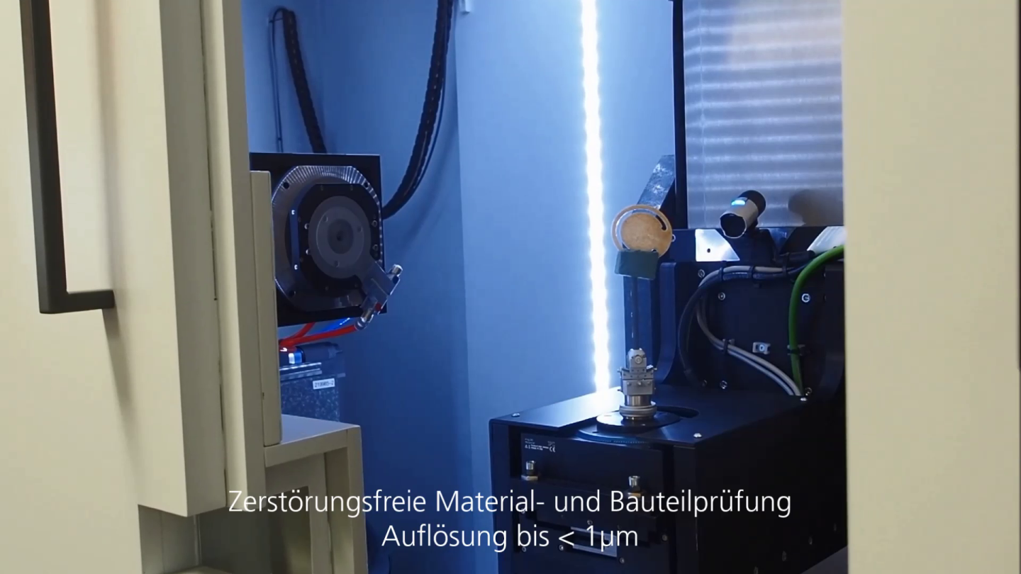
In-situ CT allows taking several consecutive images to dynamically track the behavior of the object while it is subjected to an external load - for example, a mechanical, thermal, or corrosive load.

We are able to subject material samples to a series of complementary in-situ investigations. These include tensile, compression and flexural tests, which can be performed in a temperature window between -20 and +160 °C and either linearly or cyclically.

In contrast to commonly practiced tensile or compression tests, flexure tests involve a complex loading case with simultaneous influence of tensile and compressive forces, through which more extensive information about the failure mechanisms can be acquired.

We have the capability to perform 4-point flexure tests at multiple scales:

  • Small specimens (approx. 24 x 10 mm) at high resolution (< 10 µm).
  • Larger, practical specimens (up to 150 mm x 25 mm) in accordance with DIN EN ISO 14125

Comparative testing at several scales makes it possible to visualize influences of the limited specimen dimensions for high-resolution scans and thus include them in the evaluation of failure processes. The modular design of the in-situ flexure test also allows adaptation to specimen geometries that deviate from DIN EN ISO 14125.